Hobby
Independent mathematics, AI, and machine-learning research
My hobby is independent mathematics, artificial intelligence, and machine-learning research. My current project asks a practical question: when an image is blurred, rotated, resized, poorly exposed, or corrupted by sensor noise, how does the information extracted by a computer change? I want to understand whether the pattern of those changes can help identify what went wrong during acquisition. This project continues the work begun in my University of Bristol final-year dissertation.
Retinal imaging and diabetic retinopathy
My interest in retinal imaging has a personal origin in an experience with diabetic retinopathy. It led me to take a particular interest in the reliability of retinal-image analysis.
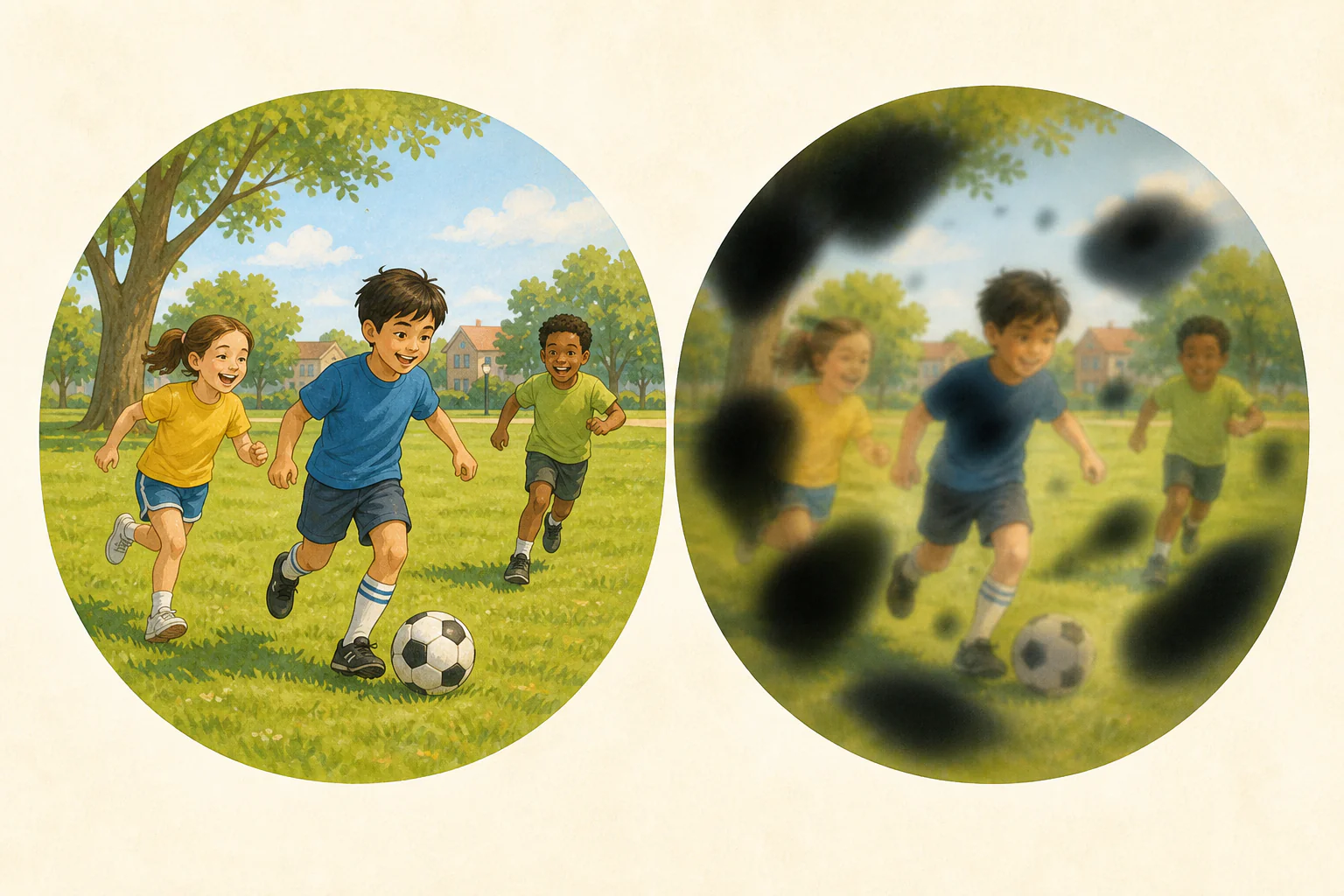
The NHS Diabetic Eye Screening Programme offers a practical setting for this question. Fundus photographs are assessed against defined feature-based criteria within a structured grading and referral pathway. Trained graders inspect the images, uncertain or referable findings receive further review, and people who may need treatment are referred for clinical assessment.
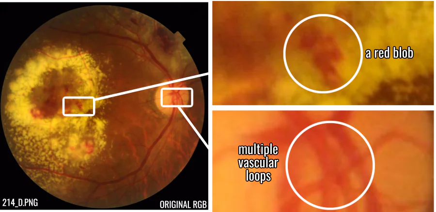
214_D from the FIVES fundus-image dataset; reused and adapted under CC BY 4.0.
As the number of people requiring screening grows, so does the grading workload. Expanding trained grading capacity also takes time. The UK National Screening Committee recently consulted on automated retinal-image analysis as a possible way to triage images before human grading. At the time of the consultation, it did not recommend introducing automated grading into the programme. A conservative role for such systems is to support trained graders and clinical teams by handling suitable routine images and flagging abnormal, uncertain, or ungradable cases for human review. The research question is what mathematical and technical assurances would be needed before that assistance could be trusted at screening scale.
The same concern with reliability has a second practical direction: more informative quality checks and fault investigation in medical or industrial imaging systems. Some faults might eventually be assessed remotely before a specialist travels onsite. This is a direction for the research, not a capability of the present work.
How the project changed direction
My original intention was to produce a topology-informed classifier for diabetic retinopathy. Classifiers of this kind already existed, but reproducing and extending established work would still have been a useful objective for a four-month undergraduate project.
The direction changed during ablation testing, when I deliberately subjected the images to individual forms of degradation. A simulated rotation of roughly three degrees was enough to drive the selected topology-based classifier close to chance performance. That raised a practical question: what value would the classifier have if a slight change in the subject’s head alignment could cause it to fail? The topological vectors were highly sensitive to rotation and other mechanical changes that required the image to be resampled. Interpolation on the pixel grid could create or destroy topological features even when the underlying subject had changed very little. My undergraduate thesis described this behaviour as grid shattering.
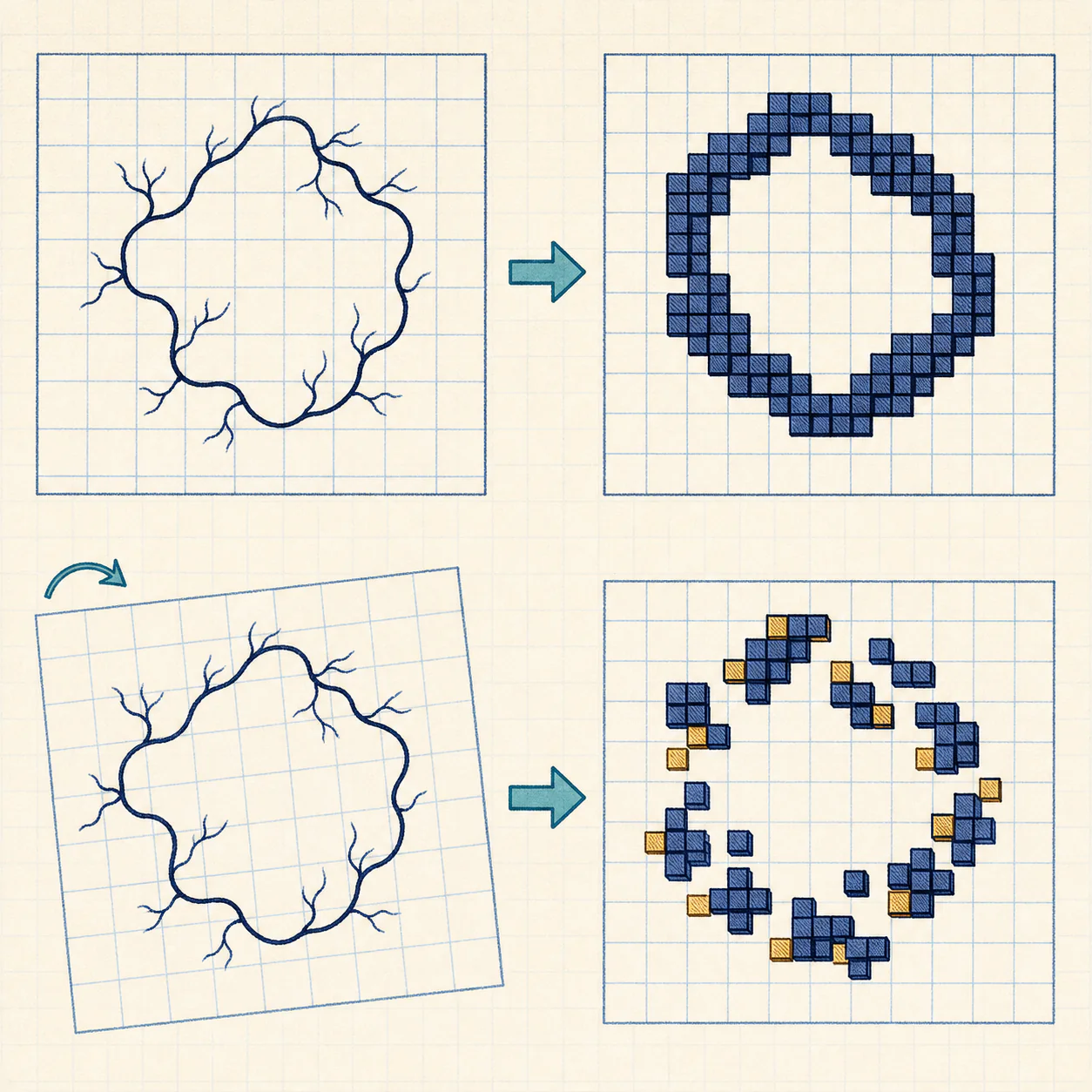
Once the ablation tests exposed this limitation, the project changed direction. Instead of treating the result as an inconvenience to the proposed classifier, I made it part of the research question and asked whether the contrasting failure behaviour could itself be useful.
In my thesis experiments, the same topological summaries were comparatively robust under several photometric changes, including exposure drift and quantisation, although not under every form of noise. Log-transformed Hu moments, which are geometric descriptors based on an image’s overall shape, showed a broadly reverse profile: they were comparatively strong under mechanical transformations but weaker under changes in intensity. Instead of asking either representation to serve as the classifier by itself, I treated them as two complementary, or “orthogonal”, controls.
I organised their contrasting failure profiles into what I called the Differential Failure Matrix. It was designed to test whether signatures associated with alignment and resampling could be distinguished from signatures associated with radiometric degradation. The final project was therefore not the classifier I had originally planned. It treated an apparent weakness of the cubical-persistence pipeline as a possible signal for a coarse fault-isolation heuristic. The code and supporting material are available in the project’s GitHub repository.
Questions left by the dissertation
The thesis was completed over four months. That was enough to establish and test the framework, but not to exhaust the questions it raised. The decomposition was descriptive and produced a suggestive empirical pattern, but it was mathematically coarse: its stability bound was conservative, and the final fault-isolation stage necessarily relied in part on empirical thresholds and heuristics. Time did not permit a deeper investigation of those mechanisms.
In a biomedical setting, a promising experimental result is not enough. The intended use, assumptions, limitations, performance, and possible failure modes of a method must be described and validated. My purpose in returning to the mathematics is to understand the underlying mechanisms more fully and to determine whether the heuristic parts of the pipeline can be replaced, constrained, or justified by stronger mathematical and empirical support. The longer-term aim is a pipeline whose assumptions and failure modes are more transparent and therefore easier to examine in technical and regulatory review. This is not a claim that the present work is a clinically validated or regulator-approved system.
Retinal imaging remains the immediate problem, and similar acquisition issues arise in industrial inspection and other computer-vision systems. My earlier experiments were confined to retinal images, so industrial applications remain a direction for further work.
Current focus
I am continuing to develop the mathematical foundations of this work, with a particular interest in making the system’s behaviour more explainable. Broadly, this means understanding why its components respond differently when an image changes, stating their assumptions and limitations more clearly, and improving how uncertainty and possible failures are communicated.
The work draws on topology, mathematical analysis, probability, and statistical machine learning. I am using these subjects to examine the pipeline from different angles rather than committing in advance to a particular theorem, model, or technical extension. The direction remains exploratory, and I will share further results when they are sufficiently developed.